Wednesday, March 29, 2017

HUMAN EMBRYOLOGY~ BY MAHEK NOOR DAUDPOTA~

HUMAN EMBRYOLOGY

       Human embryo-genesis is the process of CELL DIVISION  and cellular differentiation of the embryo that occurs during the early stages of development.
       In biological term, human development entails growth from a one-celled ZYGOTE to an adult HUMAN BEING.
       Fertilization occurs when the SPERM  cell successfully enters and fuse with an EGG CELL (Ovum).

       The genetic material of the sperm and the egg then combine to from a single cell called ZYGOTE and the germinal stage of prental development commence.
       Embryogenesis cover the first eight week of development at the beginning of the ninth week the embryo is termed  a FETUS.
The human embryology is the study of this development during first eight week after fertilization. The normal period  of gestation (Pregnancy) is nine month or 38 weeks

       The germinal stage refers to the time from fertilization through the development of the early embryo until implantation is completed in the UTERUS.
       The germinal stage takes around 10 days. During this stage, the zygote being to divide, in a process called CLEAVAGE.
       A BLASTOCYST is then formed and implanted in uterus.
       Embryogenesis continues with the next stage gastrulation, when the three germ layers of embryo from in the process called Histogenesis, and processes of Neurulation and Organogenesis follow.
       In comparison to the embryo, the fetus has more recognizable external  features and more complete set of developing organ. The entire process of embryogenesis involves coordinated spatial and temporal changes in gene expression cell growth and  cellular differentiation


       A nearly identical process occurs in species, specially among chordates.


        Step 1: a zygote is the single cell formed when an egg and a sperm cell fuse; the fusion is known as fertilization.
       Step 2: the first 12 to 24 hours after a zygote is formed are spent in cleavage – very rapid cell division
 

 Blastulation And Cell Differentiation
       Step 3: during blastulation, the mass of cells from a hollow ball.
       Step 4: cell begin to differentiate and from cavities.


        Step 5: During gastrulation the three germ layers form; the cell mass is now as gastrula.
       Step 5a: the primitive streak forms.
Step 6: The notochord is formed


Neurulation
       Step 6a: tubes from, making neurula.
       Step 6b: the notochord induces the formation of the neural plate
       Step 6c: the neural plate folds in on itself to make the neural tube and neural crest.
       Step 7: the mesoderm has five distinct categories.


Major Developmental Stages Of Embryonic Period (1-8thweek)

Time period (Week)
Major Events/characteristics

1st week

Fertilization, cleavage, morula formed (3days), blastocyst superficially implanted, zona pellucida disappears, inner cell mass(embryoblast)& outer cell mass(trophoblast) present. A cuboidal layer of hypoblast is formed.

2nd week

Blastocyst enlarged, completion of implantation, formation of amniotic cavity, yolk sac, extra-embryonic mesoderm, extra-embryonic coelom that later become chorionic cavity. Bilaminar disc (ectoderm, endoderm) is formed along with syncytiotrophoblast and cytotrophoblast. Primary chorionic villi appear.
3rd week
Appearance of:- trilaminar disc(ectoderm,mesoderm,endoderm), primitive streak(15 day), somites, interembryonic mesoderm, notochord neural tube,body cavities cardiovascular system
4th week
Body is narrow, tubular & C shaped limb buds appear, appearance of four pair of branchial arches, placenta begins to form, heart begins to beat, lens placodes and otic placodes formed, head & tail folds present . Three primary brain vesicles also formed


5th week

Head increases greatly in size, face formed, limb buds shows limbs, & upper limb become paddle shaped
6th week

Head dominant. Oral and nasal cavities confluent. Curvature of embryo diminished. Digital rays, footplate, finger & toes recognizable
7th week

Yolk sac is reduced to yolk stalk and limb differentiate rapidly.

8th week

Head nearly as large as rest of the body. Genital tubercle formed but sex difference is not obvious. Facial features are distinct. Eyes directed more anteriorly. Neck is established, digits of hands and feet seprated along with retrogression of tail.


Major Developmental Stages Of Embryonic Period (9-38th weeks)


Time period (Week)

Major Events/characteristics

9-12th  week

Rapid growth in fetal length, head still realtively large, eyes look anteriorly, eyelids fused, lanugo & nails present. Primary ossification centers appear.

13-16th  week

Further rapid growth in fetal length, head still relatively large, eyes widely separated but eyelids fused. External ears on side of head. Fetus look human and sex recognition possible.

17-20th  week

Lanugo covers entire body. Venix caseosa present. Hairs present on head mother Quickening brown fat forms. Eyebrows and eye lashes present.
21-25th  week

Skin wrinkled and red, head still relatively large, face child like, fingers & toe nail develop. Interalveolar walls of lungs secrete surfactant(24th week)

26-29th week

Skin wrinkles lost due to subcutaneous fats, eyelids open, hairs on head longer, CNS matured and controlled breathing movement primitive alveoli formed.

30-34th week

Skin is pink & smooth, langugo hairs has disappeared from face. Vernix caseosa thick. Nails reach end of fingers.

35-38th week

Fetus fully developed, lanugo hairs disappear, nails project beyond ends of finger and toes. Testis reachs scrotum.

266 days from conception  

Birth


Friday, March 24, 2017

The Muscular System.......BY Aveenash

                  THE MUSCULAR SYSTEM


OUR MUSCLES

  • Enable us to move our body parts.
  • Give us to our individual shape.
  • Protect and keep in place our abdominal organs.
  • Enable to us good posture.
  • Help in our circulation of blood.
  • Generate body heat when contract.
  • .
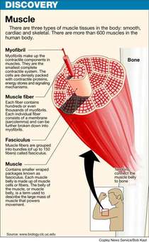
  • TYPES OF MUSCLES

  • Skeletal muscles

  • Smooth Muscles

  • Cardiac Muscles


  • 1: Skeletal or Voluntary Muscles:

  • They are under our conscious control.
  • Attached to skeleton by Tendons.
  • Causes movement of bones at joints.
  • they are fatigue.
  • Perform every day action such as: walking, runing, jumping.
  • The major Skeletal Muscles:

  • Deltoid, Biceps, Abdominals (4 muscles), Quadriceps (4 muscles), Pectorals, Latisimus dorsi, Trapizius, Triceps, Gluteals (3 muscles), Hamstrings (3 muscles), gastrocnemius.


  • 2: Smooth or Involuntary Muscles:

  • These Muscles work automatically- they are not our under consciuos control.
  • Example: Muscles of  Digestive System. 
  •  
  • 3: Cardiac Muscles:

  • Cardiac muscles are special type of involuntary muscles.
  • It is only foundst in Heart.
  • It contracts regularly, continuosly  and without tiring.
  • It works automatically but is under constant Nervous and Chemical control.
  • .


  •  HOW DO OUR MUSCLES WORK?

  • There are 3 main types of muscular contraction:
  • Isotonic and Concentric

  • Isotonic and Eccentric

  • Isometric

  • Isotonic and Concentric: Our muscles shorten as the contract.

  • Example: The bicep during a pull-up.
  • Most Sporting Movements are this type.
  • Isotonic and Eccentric: Our muscles lengthen as they contract  under tension.

  • Example: The bicep when we lower down from a pull-up.


  • Isometric:  Our muscles stay the same length as they contract.

  • There is no movement , so the ends of muscles stay the same distance apart.
  • Example: Our Shoulder Muscles during a tug of war.
  • .
  • MUSCLES CAN WORK AS:

  • Flexors: Contracting to bend our joints.
  • Extensors: Contracting to straighten joints
    .
  • Prime moovers (Agonists): Contracting to start a movement.
  •  
  • Antagonists: Relaxing to allow movement to take place.
  • Fixators: contracting to give the working muscles a firm base.
  • Synergists:  Stablising the area around the prime mover  and fine tuning our movement.
  •            MOVEMENT OF MUSCLES:

  • Origin: The attachement of muscles to the bone that remains stationary.
  •    
  • Insertion: The attachement of muscle to the bone that moves.
  • Belly: The fleshy part of muscle between the tendons of origin and/ or insertion..
  •          Muscles Disorders, Diseases and Injuries:


  • 1: CRAMPS: 

                       Involve sudden and violent muscles contractions. A person may experience painful cramps of certain skeletal muscles and smooth muscles.

  • 2: MUSCLE SORENESS/Muscle strain:  

                    Hard muscular may cause skeletal muscles become sore. In severe cases . the soreness may last up to four days. The muscle soreness may be due to the damage in muscle and connective tissue.

  • 3: Muscle Diseases/Disorders: 

  • a: Atrophy:

                  Diseases and damages the Brain or nerves that stimulate muscles.


  • b: Muscular dystrophy:  

                 Is a serious disease that directly affects muscles. It weakness the skeletal  muscles.

    BY: Aveenash 

Thursday, March 23, 2017

HUMAN SKIN--- --Rizwan

                                  HUMAN SKIN


  • The Human Skin is outer covering of body. In Human it is largest organ of integumentry system.
  • DERMETOLOGY study of Skin.

  • Skin with a total area of about 20 square feet.
  • Human skin is simillar to that most of mammals, though nearly all Human skin is covered with hair follicles.
  • Skin has a mesodermal cells, Pigmentation , such as melanin provided by melanocytes, which absorb some of potentially dangerous UV in Sunlight. It also contain DNA repair enzyme that help reverse UV damage such that People lacking these genes for these enzymes suffer high rate of skin cancer.
  • The skin is largest organ of human body, for average adult human the skin has a surface area of b/w 1.5-2.0 square meter. The thickness of skin varies considerably over all parts of body, Men and Women, Young and Old. An example is the skin of forearm which is n average 1.3mm in male and 1.26 in female.
  • The average square inch (6.5 cm2) of skin holds sweat glands, 20 blood vessels, 60,000 melanocytes and more than 1000 nerve endings.
  • SKIN HAS A THREE LAYERS

  • EPIDERMIS Layer

  • DERMIS Layer

  • HYPODERMIS Layer


  • 1. EPIDERMIS layer:- Epidermis layer is outer most layer of skin, provides a water proof barier and create our skin tone.
  • Epidermis contain no blood vessels (AVASCULAR) and cells in deepest.
  • the main type of cells which make up epidermis are Markel cells, Keratinocytes, with Melanocytes cells are present.
  • Epidermis is divided into FIVE sub-layers or strarta.
  • 1: Stratum Corneum
  • 2: Stratum Lucidum
  • 3: Stratum Granulosum
  • 4: Stratum Spinosum
  • 5: Stratum Germinatium (also called stratum basale).

  •  
  • 2: DERMIS Layer:- It is a true Skin made of Connective tissue and is a VASCULAR.

  • It is a thicker inner layer of skin.
  • matted masses of Connective tissue, Elastic Fibers, Nerve endings, Muscles, Hair follicles, Oil and Sweat glands.
  • Dermis contain lots of sensory cells (heat, cold, pain, pressure).
  • 3: HYPODERMIS Layer:- Subcutaneous. Attaches integument to muscle.

  • It is a loose Connective tissue and FAT of body's store.
  • Absorbs shocks. 
  • .
  • FUNCTIONS OF  SKIN

  • Cover-protects from germs, dehydration, injury.
  • First line of Defense.
  • Regulate body temperature.
  • Manufactures Vitamin D.
  • Site of many Nerve endings.
  • Temporary storage of Gulucose, fat, water and salt.
  • Protect from UV Radiations.
  • .
  • ONE square cm of skin contains:

  • 3,000,000 cells.
  • 10 Hairs.
  • 1 Yard of blood vessels.
  • 4 Yards of nerves.
  • 700 Sweat glands.
  • 200 nerve endings to record pain.
  • 3000 Sensory cells at the end of nerve fiber.
  • .
  • DISEASE OF SKIN:

  • ACENE:- A common and Chronic disrder of sebaceous glands.

  • ATHLETE'S FOOT:-  Fungal infection of  the epidermis.

  • DERMATITIS:- A nonspecific inflamation of skin.

  • PSORIASIS:- Patches of raised. Redish skin,

  • SKIN CANCER:-
  • Associated with UV light.


 BY: RIZWAN U-DIN CHANNA

Thursday, March 16, 2017

BONES OF SKULL

                     '' SKULL''

The skull is a bony structure that supports the face and forms a protective cavity for the brain. It is comprised of many bones, formed by intramembranous ossification, which are joined together by sutures (fibrous joints). These joints fuse together in adulthood, thus permitting brain growth during adolescence.

''BONES OF SKULL''

The bones of the skull can be divided into two groups: those of the cranium (which can be subdivided the skullcap known as the calvarium, and the cranial base) and those of the face.

The Cranium


The cranium (also known as the neurocranium), is formed by the superior aspect of the skull. It encloses and protects the brain, meninges and cerebral vasculature.
Anatomically, the cranium can be subdivided into a roof (known as the calvarium), and a base:
  • Calvarium: Comprised of the frontal, occipital and two parietal bones.
  • Cranial base: Comprised of six bones – the frontal, sphenoid, ethmoid, occipital, parietal and temporal bones. These bones are important as they provide an articulation point for the 1st cervical vertebra (atlas), as well as the facial bones and the mandible (jaw bone).

The Face


The facial skeleton (also known as the viscerocranium) supports the soft tissues of the face. In essence, they determine our facial appearance.
It consists of  14 individual bones, which fuse to house the orbits of the eyes, nasal and oral cavities, as well as the sinuses. The frontal bone, typically a bone of the calvaria, is sometimes included as part of the facial skeleton.
The facial bones are:
  • Zygomatic (2) – Forms the cheek bones of the face, and articulates with the frontal, sphenoid, temporal and maxilla bones.
  • Lacrimal (2) – The smallest bones of the face. They form part of the medial wall of the orbit.
  • Nasal (2)  – Two slender bones, located at the bridge of the nose.
  • Inferior nasal conchae (2) – Located within the nasal cavity, these bones increase the surface area of the nasal cavity, thus increasing the amount of inspired air that can come into contact with the cavity walls.
  • Palatine (2) – Situated at the rear of oral cavity, and forms part of the hard palate.
  • Maxilla (2) – Comprises part of the upper jaw and hard palate.
  • Vomer –  Forms the posterior aspect of the nasal septum.
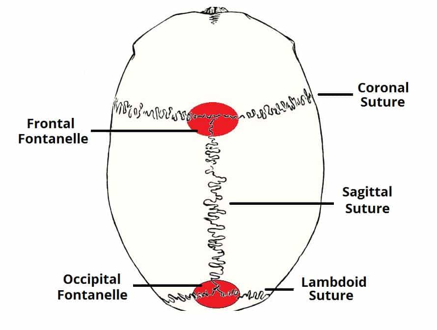
     Fig 1.2 - The major fontanelles and sutures of the skull
  • Mandible (jaw bone) – Articulates with the base of the cranium at the temporomandibular joint (TMJ).
  • Sutures of the Skull

  • Sutures are a type of fibrous joint that are unique to the skull. They are immovable, and fuse completely around the age of 20.
  • Sutures are of clinical importance, as they can be points of potential weakness in both childhood and adulthood.  The main sutures in adulthood are:
    • Coronal suture which fuses the frontal bone with the two parietal bones.
    • Sagittal suture which fuses both parietal bones to each other.
    • Lambdoid suture which fuses the occipital bone to the two parietal bones.
    In neonates, the incompletely fused suture joints give rise to membranous gaps between the bones, known as fontanelles. The two major fontanelles are the frontal fontanelle (located at the junction of the coronal and sagittal sutures) and the occipital fontanelle (located at the junction of the sagittal and lambdoid sutures).
  • Fig 1.2 - The major fontanelles and sutures of the skull
  • Clinical Relevance: Cranial Fractures


  • The majority of skull fractures result from blunt force or penetrating trauma, and can produce numerous signs and symptoms. The clinical features may be obvious, such as visible injuries and bleeding. There are also subtle signs of fracture, such as clear fluid draining from the ears and nose (cerebrospinal fluid leak indicative of base of skull fracture), poor balance and confusion, slurred speech and a stiff neck.
    There are certain areas of the skull that are natural points of weakness:
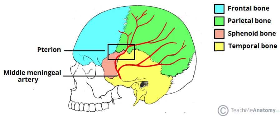
    • The pterion: a ‘H-shaped’ junction between temporal, parietal, frontal and sphenoid bones. The thinnest part of the skull. A fracture here can lacerate an underlying artery (the middle meningeal artery), resulting in a extradural haematoma.
    • Anterior cranial fossa: Depression of skull formed by frontal, ethmoid and sphenoid bones.
    • Middle cranial fossa: Depression formed by sphenoid, temporal and parietal bones.
    • Posterior cranial fossa: Depression formed by squamous and mastoid temporal bone, plus occipital bone.
    Fig 1.3 - Lateral view of the skull, showing the path of the meningeal arteries. Note the pterion, a weak point of the skull, where the anterior middle meningeal artery is at risk of damage.
    Fig 1.3 – Lateral view of the skull, showing the path of the meningeal arteries. Note the pterion, a weak point of the skull, where the anterior middle meningeal artery is at risk of damage.
  • Types of Fractures

  • There are four major types of cranial fracture:

  • Depressed – A fracture of the bone with depression of the bone inwards. They occur as a result of a direct blow, causing skull indentation, with possible underlying brain injury.
    Linear  –  The simple break in the bone, traversing its full thickness. They have radiating (stellate) fracture lines away from the point of impact. The most common type of cranial fracture.
    Basal skull – Affects the base of the skull. They characteristically present with bruising behind the ears, known as Battle’s sign (mastoid ecchymosis) or bruising around the eyes/orbits, known as Raccoon eye’s.
    Diastatic – A fracture that occurs along a suture line, causing a widening of the suture. They are most often seen in children.Fig 5.0 Skull showing depressed fracture of the frontal bone, with linear fracture marked A

    Clinical Relevance: Facial Fractures

  • Facial fractures are common and generally trauma related, i.e. road traffic collisions, fights and falls. They are often associated with clinical features such as profuse bleeding, swelling, deformity and anaesthesia of the skin. The nasal bones are most frequently fractured, due to their prominent position at the bridge of the nose.
    maxillofacial fracture is one that affects the maxillae bones. This requires a trauma with a large amount of force. Facial fractures affecting the maxillary bones can be identified using the Le Fort classification, depending on the bones involved, ranging from 1 to 3 (most serious).
  • Fig 1.4 - Le fort classification of maxillary fractures
  • BY: BILAL KHAN